Cystic Fibrosis: new image processing methods accelerate studies on drugs
A research project on cystic fibrosis, halfway between medicine and computer science, appears promising in increasing the chances of fighting this genetic disease.
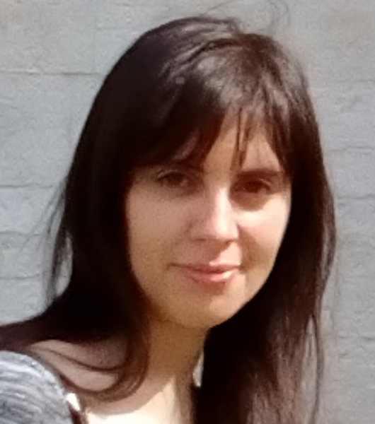
Paola Lecca, physicist and researcher at the School of Computer Science and Technology of the Free University of Bozen-Bolzano is developing advanced statistical methods for processing image data to analyze organ volume growth in medical images. Her work will speed up studies on drugs. Bioinformatics is an increasingly important research area for the School of Computer Science and Technology.
Cystic fibrosis, also known as mucoviscidosis, is a severe congenital metabolic disease caused by a mutation in the CFTR gene. A medical team from the University of Verona is focusing on the use of intestinal organoids to develop new drugs to treat this mutation.
An organoid is a simplified and miniaturized version of a three-dimensional organ produced in vitro, which has realistic microanatomy properties. These organoids are treated in the laboratory with new drugs and their growth is meticulously recorded through video microscopy. The effect and effectiveness of drugs are determined by the change in the shape of the organoid. Organoid growth is anisotropic (i.e. uneven), which is why researchers need a large number of video images to make accurate volumetric calculations.
A real bioinformatics challenge, which the School of Information Sciences and Technologies of the Free University of Bolzano and Trento-based Fondazione Bruno Kessler (FBK) are facing with the CORVO project (acronym for Computing ORganoid’s VOlume). This will shortly provide the mathematical basis that will allow us to identify abnormal changes in the volume of organoids in medical images related to cases of cystic fibrosis.
CORVO is also the name given to the software for organoid recognition in medical images that was developed in collaboration with the FBK Technologies for Vision research group.
The CORVO software analyzes temporal sequences of three-dimensional images of physiological solutions in which organoids are immersed. Through a process called segmentation, CORVO identifies the regions occupied by the organoids in each image and calculates their volumes. This procedure, carried out by FBK researcher Michela Lecca, is totally automated, but thanks to a user interface specifically designed for use by health professionals, it allows the latter to interact with the software, for example to select organoids believed to be of specific clinical interest. One of the advantages of CORVO is therefore its ease of use, since it does not require any specific knowledge of image processing by the user. Additionally, CORVO integrates a statistical analysis of the temporal variations in organoid volume that allows to sort out the different reactions of the organoids to the different pharmacological treatments.
“Bioinformatics is strategic for our School, as it shows the importance of information technology for medicine”, said the dean, prof. Claus Pahl. “I have developed a mathematical method based on advanced statistical techniques that allows us to identify, in the medical settings, which organoids develop while maintaining a spherical shape and which ones by adopting irregular shapes”, explained researcher Paola Lecca. “Intestinal organoids could be described as balloons that can be filled with water through many crevices: in people with cystic fibrosis, these CFTR channels are defective. Since water intake is largely dependent on the CFTR channels being open, organoids from healthy individuals have a full and round appearance due to regular activity while regularly working; on the contrary, the organoids of sick patients appear collapsed, smaller, and not spherical. Regular growth of the organoid in response to a drug is an indication of the drug’s effectiveness. The reduction in size and collapse of the organoid, on the contrary, are indicators that the drug is not effective. This discrimination through volume calculation would not be possible without the help of software and algorithms based on solid mathematical principles. That’s what I’m working on now”.
The experimental data for the study were produced by the cystic fibrosis applied research laboratory of the Department of Medicine (General Pathology division) of the University of Verona coordinated by Claudio Sorio and with the doctors of the “Azienda Ospedaliera Integrata” Cystic Fibrosis Center of Verona (Paola Melotti).
Paola Lecca currently carries out research activities in the team of faculty members Bruno Carpentieri (LACS Lab) and Diego Calvanese (Smart Data Factory) of the Free University of Bozen. Originally from Trento, Michela Lecca studied theoretical physics at the University of Trento, where she later earned a PhD in computer science and telecommunications. This year, she has already presented her computational model at the IEEE Symposium on Computational Intelligence and Bioinformatics and Computational Biology conference.
cs unibz/FBK
